

A team of MIT researchers has developed a revolutionary new imaging technique that allows scientists to observe up to seven different molecules at once inside living cells. This new method, described in a study published today in Cell, will give researchers unprecedented insight into the complex molecular signaling networks within cells.
According to a November 28 announcement, the technique uses fluorescent reporter proteins that turn on and off, or “blink,” at different rates. By imaging cells over time and computationally extracting each fluorescent signal, researchers can track the changing levels of multiple target proteins simultaneously.
“There are many examples in biology where one event triggers a long cascade of downstream events, which then causes a specific cellular function,” said lead author Dr. Edward Boyden, the Y. Eva Tan Professor of Neurotechnology at MIT. “This is arguably one of the fundamental problems in biology, and so we asked ourselves: Could you just watch this happen?”
Previously, typical fluorescence microscopes were limited to distinguishing perhaps two or three colors, limiting scientists to seeing only a tiny portion of the overall activity. By exponentially increasing the number of molecular signals they can visualize, Dr. Boyden’s team overcame a major obstacle.
This approach could be revolutionary for elucidating phenomena such as cell aging, cancer metastasis, learning and memory in the brain, and more by revealing how signal networks interact. The researchers have already demonstrated this in cell division cycles and plan to next study things like nutritional response, changes in gene expression and neuronal signaling.
“All of these phenomena could be thought of as representing a general class of biological problems, where a short-term event – like eating a nutrient, learning something, or contracting an infection – generates a long-term change,” Dr Boyden explained.
The key is to use fluorescent markers that flash at set rates, then computationally dissociate their signals after viewing the cells for a few minutes or even days. The team continues to work to expand its fluorescent palette even further. Importantly, implementation is simple on basic optical microscopes already ubiquitous in laboratories around the world.
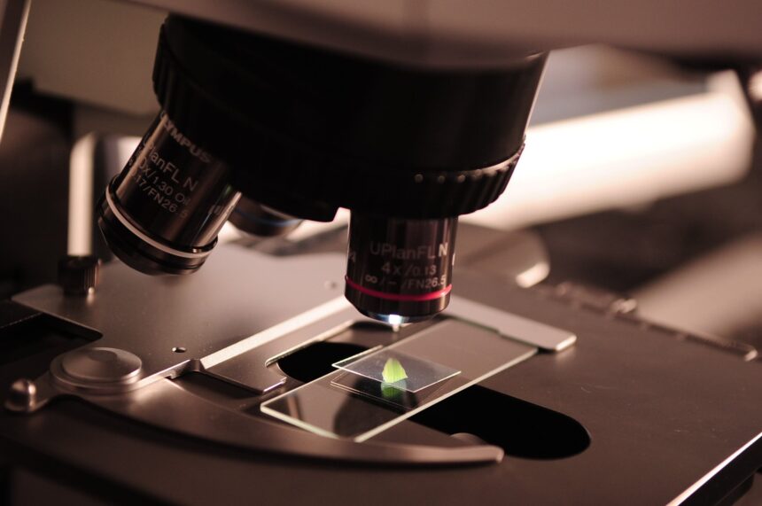
Featured Image: Pexels